
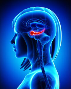
(Duke University School of Medicine, Shantell M. Kirkendoll) — Based on brain images and memory tests, the Duke study published Nov. 27 in the Journal of Neuroscience reveals a finely tuned memory dance choreographed by the hippocampus, the brain’s memory center.
The hippocampus doesn’t store detailed object information. Instead, it influences how the cortex stores such information that would be needed later, ensuring that memories are coded in a way that meets future needs and tasks.
This research can help identify specific brain regions that can be stimulated to bring back healthy brain activity in individuals experiencing memory decline, such as Alzheimer’s disease.
“What is exciting about this study is that it shows that the details supporting memory processes are fundamentally driven by the interaction between the hippocampus and the cortex, but not by each region independently,” said senior author of the study Simon W. Davis, PhD, an assistant professor in the Department of Neurology at Duke University School of Medicine and a member of the Center for Cognitive Neuroscience.
“This is a much more contemporary way of seeing the brain,” he said.
The hippocampus is believed to help in forming episodic memories by connecting pieces of information stored in the cortex. However, the exact process by which the hippocampus and cortex work together to store memories is not entirely clear.
“The study suggests that the strength of interactions between the hippocampus and other brain regions during memory encoding can predict subsequent memory,” said lead study author
Shenyang Huang, a graduate student at Duke University.
To better understand how brain regions process features of objects, researchers used functional MRI, a brain imaging technique, to observe 19 study participants as they tried to memorize images of real-world objects like pizza, birds, and clothing.
They later tested the participants on their memory of both the concept of the objects and the specific details.
Their findings revealed that certain brain regions exhibited a heightened sensitivity to visual characteristics, while others were more attuned to semantic, or meaning-related, information.
The hippocampus plays a critical role in adjusting the memory impact of cortical representations that are most relevant to future memory needs, or what authors referred to as “transfer-appropriate representations.”
In the realm of remembering how things look, the strength of visual representations in the ventromedial occipital cortex is crucial, coordinated with hippocampal activity and information patterns during encoding.
Similarly, when it comes to grasping the meaning or concept of objects in subsequent conceptual memory, the strength of semantic representations in the left inferior frontal gyrus and angular gyrus is key.
While new medications offer hope for those with age-related memory disorders, researchers at Duke are exploring alternative interventions including transcranial magnetic stimulation (TMS).
“As we search for reliable targets for TMS, it’s important to emphasize cortical targets that link to the hippocampus in an informed matter, so they can boost meaningful conversations in the brain,” said Davis, who also has appointments in the Department of Pathology and the Department of Psychiatry and Behavioral Sciences at the Duke School of Medicine, and in the Department of Psychology and Neuroscience at Duke.
Davis and colleagues gained support from the National Institutes of Health to explore how older adults with memory impairments may benefit from individualized magnetic brain stimulation.
“The efficacy of that intervention—and our understanding of why it works—will be based on studies like this showing how cortical-hippocampal communication forms strong memories,” Davis said.
Additional authors: Cortney M. Howard and Roberto Cabeza of Duke University; Mariam Hovhannisyan of University of Arizona and Maureen Ritchey of Boston College